Mon-Fri 09:00-18:00      
Tashkent city, Yunusabad district, st. Okil Ota, 15
Request a call

Shop

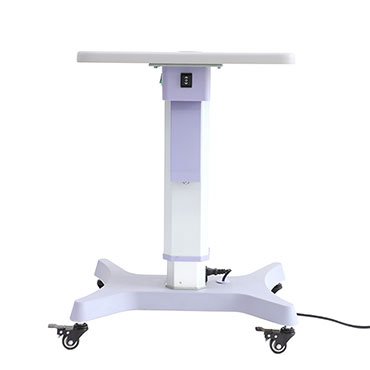
Электроподъёмный стол Hongdee HD-18AE

Ophthalmic Motorized Table Hongdee HD-18AE

Manufacturer: Hongdee (Beijing Hongdee Corporation), China

Description:
Electric motorized instrument table — the entry model of the HD-18 series. The 400 × 480 mm tabletop seats a slit lamp, auto refractometer or lensmeter; one power outlet beneath the tabletop; plastic base, no drawer.

Application:
Holding tabletop ophthalmic instruments with electric height adjustment to the patient.

Офтальмологический операционный стол модель Comfort 5.0

Ophthalmic operating table model Comfort 5.0

The Stephen H Comfort 5.0 chair from Gardhen Bilance is designed to ensure adequate comfort for patients of all sizes during surgery and medium and long-term therapies. Thanks to the interchangeability system of the headrests and the numerous accessories available, this medical device is particularly suitable for minor surgery interventions.

Офтальмологический операционный стол Neotech ОТ-2001

Ophthalmic operating table Neotech ОТ-2001

Altitude range min./max. Overall length: 580 mm / 810 mm
Overall width: 1860 mm
Lifting speed: 610 mm
Back section Load capacity: Electric cylinder
Input voltage: 135 Kgs
Gross 230 V 50/60 Hz. , 110 V 50/60 Hz.: 118 kgs
Altitude range min./max. Total length: 135Kgs

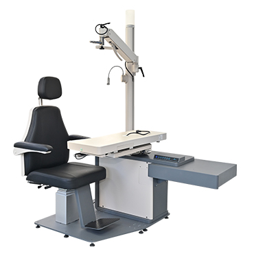
Рабочее место офтальмолога Hongdee HD-100E

Ophthalmic Refraction Unit Hongdee HD-100E

Manufacturer: Hongdee (Beijing Hongdee Corporation), China

Description:
Refraction chair unit — patient chair plus instrument table — from Hongdee’s combined workstation range. The sliding tabletop seats two instruments together; compact footprint for space-limited rooms.

Application:
Ophthalmologist/optometrist workstation setup; left-handed or right-handed configuration available.

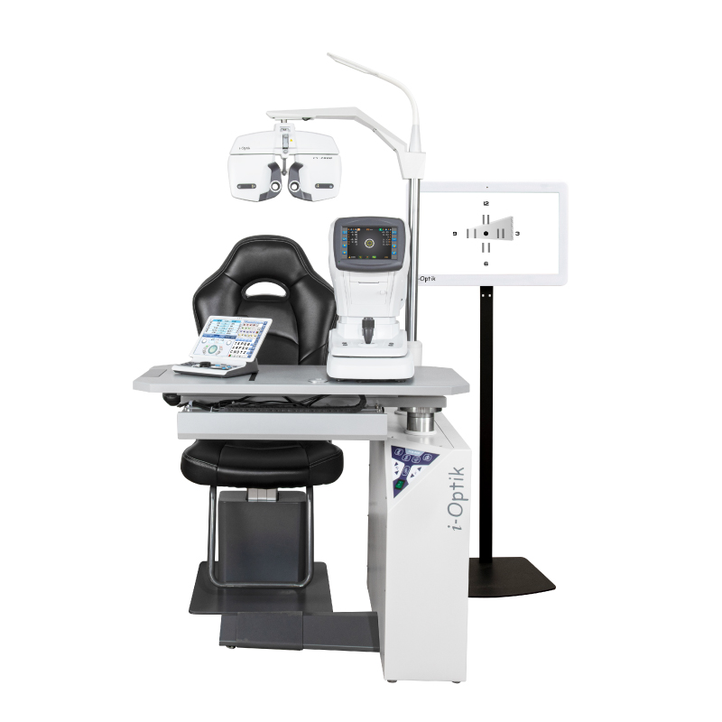
Рабочее место офтальмолога i-Optik TCS-900

Ophthalmic Refraction Unit i-Optik TCS-900

Manufacturer: i-Optik (Ningbo Ming Sing Optical R&D), China

Description:
Combined workstation: patient chair and instrument table in one unit. The phoropter arm is motorized for lifting and rotation; the table top opens to 90 degrees and slides to hold two instruments at once.

Application:
Refraction room setup: auto ref/keratometer, slit lamp and phoropter on a minimal footprint, with a motorized patient chair.

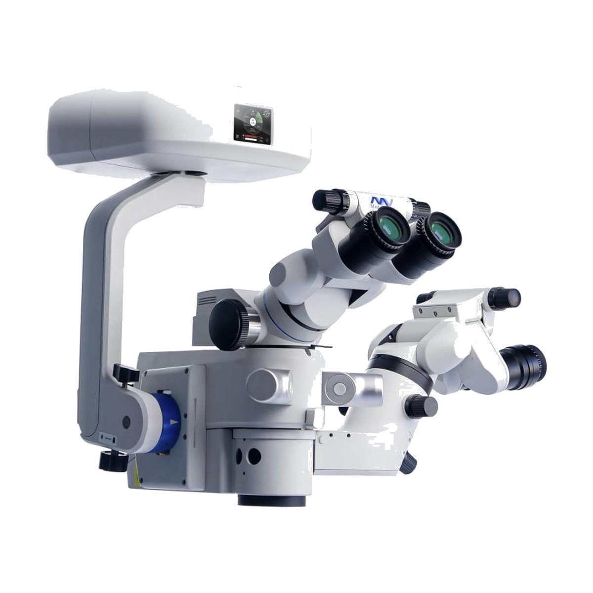
Ophthalmic Surgical Microscope MediWorks SM621

Ophthalmic Surgical Microscope MediWorks SM621

Manufacturer: MediWorks, China

Description:
The SM621 is an ophthalmic surgical microscope with motorised apochromatic optics and a 1:6 zoom ratio. Coaxial multi-directional stereoscopic LED illumination with independently adjustable red reflex delivers a bright, stable reflex. Control is via a 14-function wireless foot panel and a multifunctional touch-screen display. The microscope holds CE, NMPA and FDA approval; a 4K digital video module, the 600B non-contact wide-angle viewing system and the 600A stereoptic inverter are optional.

Application:
Anterior segment microsurgery — cataract phacoemulsification and IOL implantation, where a stable red reflex is critical. With the optional 600B system and 600A inverter it extends to vitreoretinal surgery.

Офтальмологический ультразвуковой аппарат для A/B сканирования aviso с подставкой + UBM 50 HZ

Ophthalmic ultrasound apparatus for aviso A/V scanning with support + UBM 50 Hz

The software allows you to:
– Record sequences of images up to 10 seconds long. For more information, see Cineloop,
– view images recorded during previous scans,
– save the images in the patient’s file in the computer database,
– print an official report including user data, patient data and up to 6 selected images.

Before you begin to retrieve an image, the user must do the following:

– install the system, connecting the PC and sensors to the AVISO module,
– turn on the PC that comes with the software
AVISO software,
– configure the system,
– enter user and patient data,
– make a B-scan image and display the Cineloop sequence,
– save the selected B-scan image,
– create an official report with many images, save and
print it on your printer.

DGH Scanmate Flex АБ-скан (10/20 МГц)

Ophthalmic Ultrasound DGH Scanmate Flex A/B-Scan (10/20 MHz)

Manufacturer: DGH Technology, USA

Description:
Portable ophthalmic ultrasound platform that plugs into the USB 2.0 port of a Windows computer — no dedicated console. A/B configuration: A-Scan 10 MHz for biometry and B-Scan 12.5/20 MHz for posterior-segment imaging.

Application:
Ultrasound biometry (axial length, ACD, lens thickness) and B-scan imaging of retina, vitreous, choroid and optic nerve. The platform can be expanded with a UBM probe.

DGH Scanmate Flex АБ-скан + UBM (10/20/50 МГц)

Ophthalmic Ultrasound DGH Scanmate Flex A/B-Scan + UBM (10/20/50 MHz)

Manufacturer: DGH Technology, USA

Description:
Portable ophthalmic ultrasound platform that plugs into the USB 2.0 port of a Windows computer — no dedicated console. Three-modality configuration: A-Scan 10 MHz (biometry), B-Scan 12.5/20 MHz (posterior segment) and UBM 35/50 MHz (anterior segment, ClearScan water-path).

Application:
Ultrasound biometry (axial length, ACD, lens thickness), B-scan imaging of retina, vitreous and optic nerve, ultrasound biomicroscopy of the angle and ciliary body.

Офтальмологический ультразвуковой аппарат для A/B сканирования

Ophthalmological ultrasound machine for A/B scanning

Advantages of A/B scan CAS-2000BER:

🔹 Ability to record photos and videos
🔹 Provides high image quality with great processing capabilities
🔹 Frame-by-frame viewing of video clips allows you to choose the optimal image
🔹 Easy to use for measuring distances and angles in an image
🔹 It is possible to maintain a database of patients with libraries of images and video clips and printed examination results

Офтальмоскоп BXa-RC

Ophthalmoscope BXα-RC

Better balanced reduction of corneal reflex and bright fundus image

Features:
– Corrective lens from -36 D to +35 D in 1 D increments for precise focusing
– Unique polarizing filter for observing natural fundus images
– Original Neitz optical systems for minimal corneal reflex
– Dust curtain to prevent foreign objects from entering
– Battery power supply. No need to replace battery

Офтальмоскоп YZ11D

Ophthalmoscope YZ11D

A halogen lamp with uniform illumination displays the real colors of the fundus tissues. There is an intelligent system, convenient for mobile diagnostics. The device is equipped with a special charger that provides fast and convenient recharging.

Оптический биометр + топограф Topcon MYAH

Optical Biometer and Corneal Topographer Topcon MYAH

Manufacturer: Topcon Healthcare, Japan

Description:
Compact optical biometer and corneal topographer for myopia management programmes. Axial length via low-coherence interferometry (LCI), corneal topography (24-ring cone, 100,000+ analyzed points), white-to-white, dynamic pupillometry, meibomian gland imaging.

Application:
Myopia progression monitoring with visit-to-visit trend reports; contact lens fitting including Ortho-K databases; dry-eye assessment.

Optical Biometer MediWorks AL550

Optical Biometer MediWorks AL550

Manufacturer: MediWorks, China

Description:
The AL550 is an optical biometer built on optical low-coherence reflectometry at 1,060 nm — a strongly penetrating wavelength that keeps the measurement success rate high even in dense cataract. It integrates a 50-ring Placido disc corneal topography module analysing 125,600 data points on the anterior surface. A single measurement returns up to 22 parameters, and a full binocular examination completes in 60 seconds with full-automatic focus and voice prompts.

Application:
Pre-operative biometry for cataract and refractive cataract surgery: axial length, anterior chamber depth, lens thickness, keratometry and white-to-white for IOL power calculation with built-in Pearl-DGS (optional), Jin, SRK-T, Holladay 1, Hoffer Q and Haigis formulas. In paediatric practice it monitors myopia progression via axial length and AL/CR trend curves.

Оптический когерентный томограф Dri Oct Triton Topcon

Optical coherence tomograph Dri Oct Triton Topcon

B-scan. The large informative content of the images contributes to improving the efficiency and quality of diagnostics.

The high penetrating power of light from a frequency-modulated source provides a simple and clear visualization of such deep layers of the eye as the choroid and sclera. Another advantage of a frequency-modulated source is the ability to visualize the vitreous body and the vascular membrane equally clearly and without interference in one scan. Due to this, there is no need to waste time on separate scans of the vitreous and vascular membrane